-
home
Introduction and mission statement of our project.
-
people
Information about the PI, postdocs and students.
-
research
Description of our research, with brief introduction to our projects in computer vision, computational biology and neuroscience, and medical image computing.
-
publications
Journal and peer-review conference publications, and some abstracts.
-
collaborations
A list of our collaborations within the last five years. We acknowledge their contribution and their novelties in their fields.
-
sponsors
Federal organizations and private institutions that fund our research.
-
links
Some resources that we use and you may find useful.
-
contact
Contact information, including phone number, email, and location.
Medical Image Computing
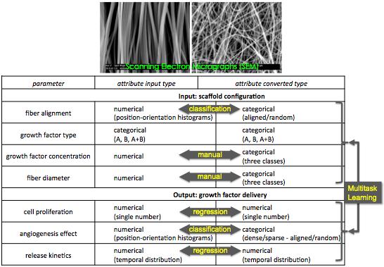
Rational design of cytokine releasing angiogenic constructs (NIH: 1 R21 EB012136-01)
(Prediction of the angiogenic effect of growth-factor delivering scaffolds)
A collaborative work with Fotios Andreopoulos (BME, U of Miami).
The development of organized vascular networks requires a series of highly specific interactions between cells, growth factors and soluble mediators. Among the various approaches (e.g. cell transplantation, surgical intervention, or drug therapy) to promote vascular regeneration, therapeutic angiogenesis based on the delivery of soluble cytokines has generated considerable interest because of its minimal invasiveness and promising pre-clinical success.
Our objective is to synthesize bioactive, three-dimensional scaffolds with patterned architecture, demonstrate that the spatial and temporal delivery of two model angiogenic growth factors has an enhanced angiogenic effect and develop models that can predict the biological effect of a growth factor releasing construct as a function of fabrication parameters.
The hypothesis is that guided therapeutic angiogenesis (i.e. patterned vascular networks)
is possible by controlling the spatial and temporal presentation of soluble mediators at the site of ischemia.
By designing nanofibrous scaffolds that direct the local gradients of angiogenic cytokines we could manipulate the proper migration of cells that presages vascular patterning.
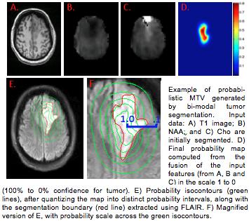
Radiation Treatment Planning of Primary Brain Tumors
A collaborative work with Radka Stoyanova (U of Miami) and the Radiation Oncology Department of U of Miami.
Radiation Treatment (RT) planning aims at delivering the highest radiation dose to the active disease while sparing normal tissue as much as possible. Conventional imagery, such as MRI and CT are used to define the target tumor's 3D shape and volume for irradiation. In many cases these provide ambiguous or limited information with regard to tumor infiltration. In contrast, 1H MR spectroscopic imaging (MRSI) has the ability to identify complex biochemical patterns associated with normal brain, tumor histopathology and changes in tissue function.
In our work, we combine MRI (T1, FLAIR) and MRSI (Choline and N-acetylaspartate marker) to generate Metabolic Tumor Volume (MTV) maps which may redefine the standard treatment volumes utilized in RT of brain tumors. Our computational framework consists of a 3D Conditional Random Field model, in which the inference is driven by boosting-based feature selection in an online learning manner.
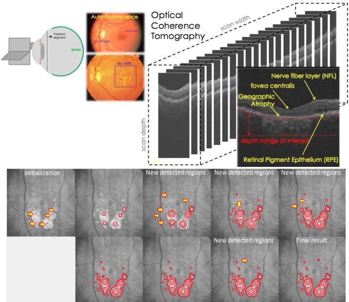
Quantification geographic atrophy in dry age-related macular degeneration of the retina
A collaboration with Bascom Palmer Eye Institute.
Age-related Macular Degeneration (AMD) has become one of the most common causes of severe irreversible vision loss in developed countries. In patients with advanced dry AMD, most of the severe vision loss results from atrophy of the retinal pigment epithelium (RPE). Confluent areas of RPE atrophy are clinically referred to as geographic atrophy (GA), which can cause legal blindness if it affects the central macula. There is currently no effective treatment for GA and there is only a rudimentary understanding of its pathophysiology. Furthermore, its visibility by standard photography depends on the degree of pigmentation present in the surrounding intact RPE. Spectral Domain Optical Coherence Tomography (SDOCT) demarcates areas of GA precisely even when it cannot be identified by photography.
For the automated quantification of the GA, we use a geometric model driven by dynamically updated probability fields. The image probability fields are estimated by our collaborative Conditional Random Field (CoCRF), which is updated during the evolution in an online learning manner: it infers class posteriors in pixels or regions with feature ambiguities by assessing the joint appearance of neighboring sites and using the classification confidence.
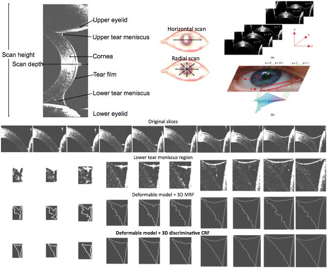
Measuring the human tear meniscus obtained with spectral-domain Optical Coherence Tomography
A collaboration with Bascom Palmer Eye Institute.
The dynamic variation of the human tear meniscus (tears around the eye lids) is very critical in visual function, maintenance of corneal integrity, and ocular comfort. The quantitative measuring of the tear menisci around the eyelids is though a challenging task. In our work, tear meniscus images are obtained with a custom-built Optical Coherence Tomography (OCT) device and are segmented using a probabilistic (Random Field-based) geometric model. Our results show that our segmentation approach successfully handles clutter and boundary ambiguities of the tear menisci, which makes our integrated system reliable for the every day medical practice.
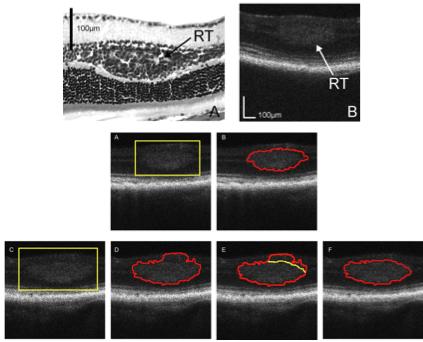
Detection of retinal tumors in the mouse model of retinoblastoma
A collaboration with Bascom Palmer Eye Institute
The retinal tumor in a mouse model has been successfully imaged using an ultra-high resolution spectral-domain optical coherence tomography (SD-OCT) designed for small animal retinal imaging. For segmentation of the tumor boundaries and calculation of the tumor volume, we developed a novel segmentation algorithm, based on active contours driven by propability fields derived from the image observations. With this algorithm we are able to obtain the tumor boundaries automatically, while the user can specify additional constraints (points on the boundary) to correct the segmentation result, if needed. The system and algorithm were successfully applied to studies on retinal tumor progression and monitoring treatment effects quantitatively in a mouse model of retinoblastoma.